So yet again I’ve taken a long hiatus from blogging – but generally that’s a good thing, as I’ve been so busy with enjoying life and getting back to things that complaining about it on my blog has taken a back seat!
At the request of several family members; here is an update on how my angiogram, which I was preparing for in my last post, went.
This angiogram took place on the 30th of December, in that odd period between Christmas and New Year that nobody really knows the purpose of. This wasn’t my first angio; it was actually my fourth. However this was the first one that I would be conscious for, and when you know you’re going to have a wire and tube inserted up the artery in your leg and threaded all the way into your brain, even an above average amount of insight doesn’t really help to calm the nerves, the anticipation and, most importantly, the curiosity.
The day approached. I was up bright and early, as I had to check in with the hospital at 8am. There was some initial confusion as to where to go – my letter had suggested I had to go to Outpatients, despite the fact that I would be an inpatient for a day. Eventually we were directed to a ward, and I was sad that it wasn’t my usual ward, Victor Horsley. Instead we were sent to, rather strangely, a ward emblazoned with “Nuffield Health, Queen Square Private Ward”. What? Thinking that we were going to have to move yet again, we entered the ward with some trepidation… only to discover that we were indeed expected in the private ward and my bed was waiting for me. What a pleasant surprise!
The private ward was plush; you truly do get what you pay for. Each bed had its own separate room, with a flatscreen TV, ensuite bathroom, wardrobe and… a MINI FRIDGE in the wardrobe! Interestingly, the bed in the private ward was exactly the same as the bed in the NHS ward. So whether you were prince or pauper, you still slept on the same mattress. I feel like there is some clever, significant saying I could make out of that but I’ll leave that to you guys to figure out. Having been on morning take before and having been the patient subjected to morning take, I knew the drill and patiently waited to have my obs done and be clerked in. An hour later, I was still waiting. And starving too, as I had to be nil by mouth from midnight. Eventually a nurse came to take my obs whilst a locum doctor from America clerked me in and took my blood. I ended up having to point out where my vein was and stabilise it, as she missed it first time and pierced through the vessel. Oh dear. Shortly after, the interventional radiologist that was going to do the procedure came to take consent from me… not only for the angiogram, but also to ask if I would be happy to do an MRI afterwards for a study he was doing. I instantly said yes, and tried to see if there was an opportunity to help. Sadly he missed the meaning of that and I only helped by contributing an image of my brain.
Yet more time passed, mostly interspersed with me napping, chatting to my boyfriend’s mom who had accompanied me or sending hospital/”omg private ward” selfies on snapchat. 12pm rolled around and I was taken downstairs to the theatres, and so the angiogram experience began. As I was taken into the theatre, I noticed on the wall the very same poster of the anatomy of the brain that I had stuck to my bedroom wall, which made me smile. My femoral artery was found (handily signposted by my scar from my previous angiograms), skin was prepped and local anaesthetic injected. Which, in hindsight, was the most painful/uncomfortable part of the whole procedure. I had requested some form of blanket as the theatre was quite cold and I had a fan heater blowing heat onto me underneath the sterile drapes. Which was amazing, until the local anaesthetic kicked in and I then had a cold triangle of skin; a very odd sensation. Satisfied that the patch of skin was numb, the catheter (4 french; roughly equivalent to 1.3mm) was inserted and threaded up my arterial tree. This again was an odd sensation; I could sort of feel it as a slight pressure in my common iliac, when it hit my abdominal aorta I could feel it less and once it went past my heart, I could feel absolutely nothing. Even though I could see the radiologist pushing and pulling quite forcefully at times, nothing could be felt. Eventually he reached my internal carotid and this is where the fun really started.
I had been warned, and read interesting stories, of the effects that the dye injected to visualise the vessels had on your brain. My handy “What to expect” booklet had told me of perhaps seeing flashing lights, hearing strange sounds, tastes; and my doctor had said the same. He was going to warn me everytime he injected the dye, after which I had to hold my breath for the duration of the contrast and X-Ray. Much to my annoyance, the screens were sort of in my left peripheral vision, but I couldn’t see them properly unless if I turned my head. Which I wasn’t allowed to. I had to stare straight up at the white box that was the tail end of the X-ray machine. But this white box provided the perfect blank canvas on which my visions grew. The first time I was so taken aback by what had appeared in front of me that I didn’t have time to analyse it before it ended. “Wow,” I told the doctor, “That is certainly an experience.” He replied saying he had heard similarly from other patients, and almost wished he could experience it too. The next time he injected, I was prepared.
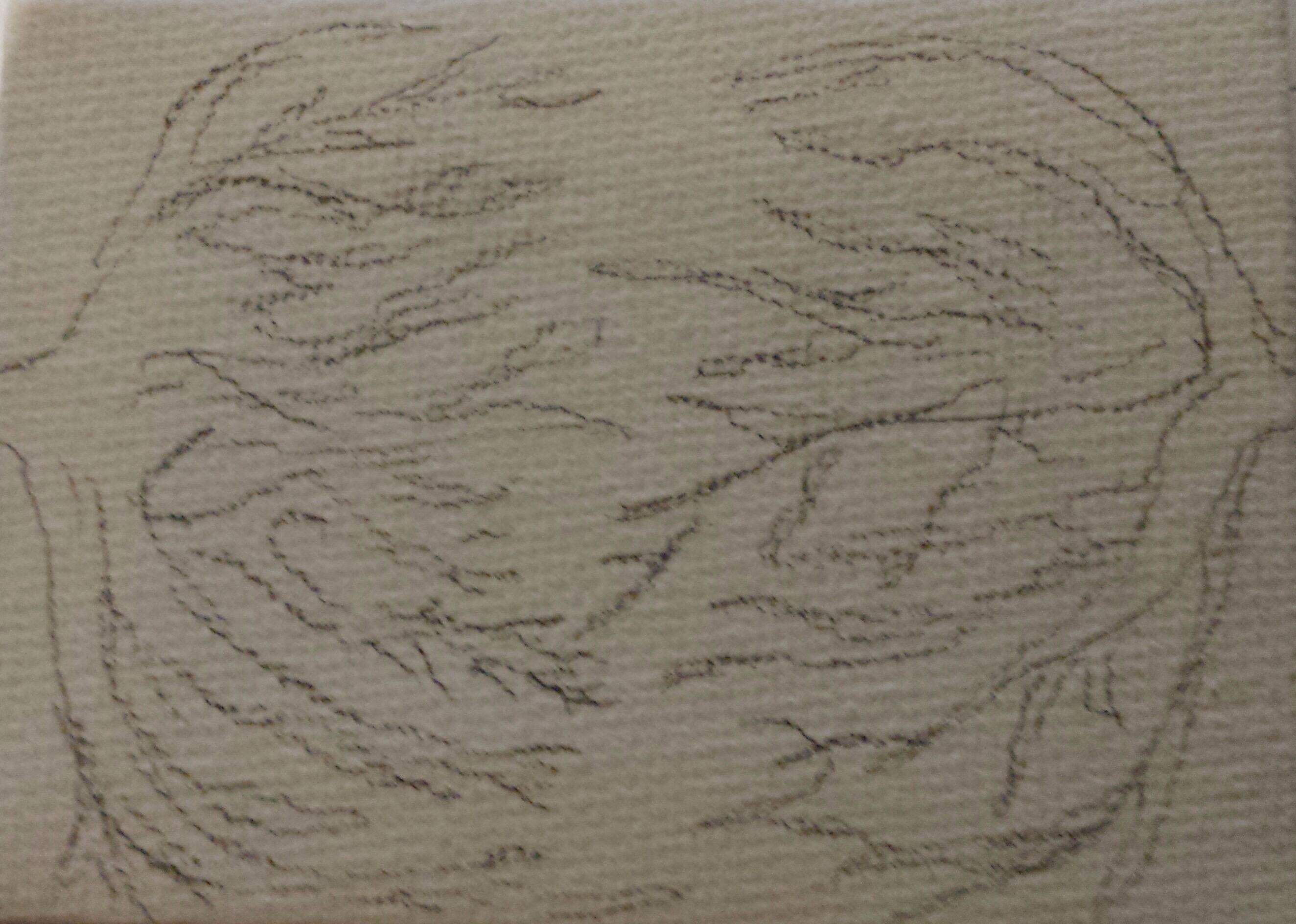
It started in the center of my vision; a white glowing square that grew until it reached the edges, then it faded out to be replaced by some white glowing, pulsating, branching structures growing from the peripherals of my vision and sprawled across back to the center. I say branch, but in all honesty my first though was I was somehow seeing my retinal blood vessel network. It was very organic; it looked like a tree in winter, like a network of vessels or a network of neurons. Whilst this was going on, it was accompanied by a soundtrack that filled my head; a crackling, popping sound and sensation very similar to having too much popping candy in your mouth… but it was in my BRAIN!
These effects happened every time dye was injected into that artery, but once the target artery was changed, the effects changed. On one injection, exactly half of my face became a temporary radiator, which changed to the other side of my face when the same artery on the other side was injected. Then a new artery was selected and I had a strange, almost metallic taste going down the back of my tongue and throat. After a few more injections the team were happy. I tried to ask them as many questions as possible but I had to hold my breath so many times I ended up staying silent. As they were wheeling me out of the theatre I manage to get a glimpse of the screens and saw Ralph in all its twisty glory. I was astounded; it was like I had been blind previously and now I could see. Ralph’s nidus was suddenly there, in full HD glory. It was like watching a YouTube video in 1080p instead of 240p resolution.
When I was in recovery and my wound was being compressed, Dr Rennie, the wonderful interventional radiologist who did my embolisation back in June, came to say hi. We discussed a lot; mostly neuroanatomy, specifically MY neuroanatomy. He was happy with the angio today; it turns out the reason why previous angio’s haven’t been clear was because the varix was occluding the main nidus and casting a shadow on it. But now that my AVM was more visible, just how complicated it really was was also more visible. It was previously thought that my AVM had one feeding artery, and one draining vein. Like most AVMs did. It turns out, Ralph has not one, but THREE feeding arteries, and was starting to recruit dural blood vessels to drain into. Which, in summary, meant that Ralph was still pretty complicated, and we still had to watch and wait.
After having a very quick MRI for the study, I was taken back to the ward. On the way back I expressed concern about missing lunch, to which a nurse assured me that seeing as I was on the private ward, if I missed lunch they would cook a new one from scratch for me. Thankfully I hadn’t missed lunch and was presented with a posh plate of tomato soup, bangers and mash, and sticky toffee pudding under…. a silver dome. Yep, my ward had silver service. And asking for some water resulted in me being given a monogrammed glass carafe of some fancy alpine, volcano-filtered still water, instead of the plastic jug of tap water that I was used to. I could see why people paid for private now – if not for the healthcare, then for the hotel like service and food!

After lunch amongst my visitors was Mr Shieff, who came to check in and update me with what Dr Rennie had already said. He mentioned that the gamma knife team would be looking at these images and come to a decision. They haven’t. I’m typing this on 18/02/15, almost 2 months later, and so far the decision has been made to give me another MRI scan.
Eventually I was discharged around 7pm. Overall the angiogram experience had been more fascinating than anything. Possibly the most irritating aspect was the part where I had to lie completely flat for 6 hours after my angiogram, to minimise complications of the wound site. That doesn’t sound bad on the surface, but try drinking tea through a straw, lying flat. And if you needed the toilet…. well, you just learn to have a very strong bladder. All those weeks spent holding my bladder on Duke of Edinburgh expeditions did come in handy after all!

Any scan images to compare progress with original “Ralph”?
Not yet! Still waiting, but I will definitely upload comparison scans when I get them 🙂
Your experience sounds a lot like the visuals you get when you’re on lsd 😀
Pingback: My 2nd second birthday | Me, myself and Ralph